 |
|
|
|
|
||||||||
|
Color
Perception And Visual Acuity
By Jeffrey W. Johnson |
||||||||
 |
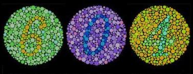
Two aspects of the human vision that you will need to have are color perception and visual acuity. Included below are two quick tests for both color and acuity: COLOR PERCEPTION: Shown above is a sample of the type of color images that you will be asked to identify by your medial examiner. In each of the above circles is a number. If you can identify the numbers of each of the circles, then chances are you have no color vision deficiencies. |
|||||||
|
Myself, I cannot see the 0 that is in the center circle. I failed to identify the color differences associated with those of the center circle and therefore failed that portion of my medical exam. The restrictions to a pilot's license that apply for such a vision deficiency are "no night flight" and "not valid for color control signal". If you have a similar problem and still have the restriction, click here to learn about the process to obtain a S.O.D.A. (Statement Of Demonstrated Ability ). Federal Aviation Regulations, according to the third-class qualifications, sec. 67.303 says: Eye standards for a third-class airman medical certificate are: (c) Ability to perceive those colors necessary for the safe performance of airman duties. ** Note: This actually means the ability to distinguish between red, green, and white lights.
|
||||||||
|
- Very bottom line = 20/10 vision - Second line up from bottom = 20/20 vision - Third line up from bottom = 20/30 vision - Fourth line up from bottom = 20/40 vision - Fifth line up from bottom = 20/50 vision - The "T" and "B" represent 20/100 vision - The "E" at the top represents 20/200 vision Federal Aviation Regulations, according to the third-class qualifications, sec. 67.303 says: Eye standards for a third-class airman medical certificate are: (a) Distant visual acuity of 20/40 or better in each eye separately, with or without corrective lenses. (b) Near vision of 20/40 or better in each eye separately, with or without corrective lenses. ** Note: if corrective lenses are required to obtain the minimal 20/40 vision, then the person is eligible only on the condition that the corrective lenses MUST be worn while exercising the privileges of an airman certificate. |
||
|
An eye examination is a battery of tests performed by an ophthalmologist, optometrist, or orthoptist assessing vision and ability to focus on and discern objects, as well as other tests and examinations pertaining to the eyes. Health care professionals often recommend that all people should have periodic and thorough eye examinations as part of routine primary care, especially since many eye diseases are asymptomatic. Eye examinations may detect potentially treatable blinding eye diseases, ocular manifestations of systemic disease, or signs of tumours or other anomalies of the brain. Color vision is the capacity of an organism or machine to distinguish objects based on the wavelengths (or frequencies) of the light they reflect, emit, or transmit. The nervous system derives color by comparing the responses to light from the several types of cone photoreceptors in the eye. These cone photoreceptors are sensitive to different portions of the visible spectrum. For humans, the visible spectrum ranges approximately from 380 to 740 nm, and there are normally three types of cones. The visible range and number of cone types differ between species.
A 'red' apple does not emit red light. Rather, it
simply absorbs all the frequencies of visible light shining on it
except for a group of frequencies that is perceived as red, which
are reflected. An apple is perceived to be red only because the
human eye can distinguish between different wavelengths.
The advantage of color, which is a quality constructed by the visual brain and not a property of objects as such, is the better discrimination of surfaces allowed by this aspect of visual processing. In some dichromatic substances (e.g. pumpkin seed oil) the color hue depends not only on the spectral properties of the substance, but also on its concentration and the depth or thickness. Color processing begins at a very early level in the visual system (even within the retina) through initial color opponent mechanisms. Opponent mechanisms refer to the opposing color effect of red-green, blue-yellow, and light-dark. Visual information is then sent back via the optic nerve to the optic chiasma: a point where the two optic nerves meet and information from the temporal (contralateral) visual field crosses to the other side of the brain. After the optic chiasma the visual fiber tracts are referred to as the optic tracts, which enter the thalamus to synapse at the lateral geniculate nucleus (LGN). The LGN is divided into two zones: the M-zone, consisting primarily of M-cells, and the P-zone, consisting primarily of P-cells. The M- and P-cells make up the principal sublayers, while each layer has a koniocellular sublayer. M- and P-cells received relatively balanced input from both L- and M-cones throughout most of the retina, although this seems to not be the case at the fovea. The koniocellular sublayer receives axons from the small bistratified ganglion cells, which directly compare the output of S-cones and L/M-cones, giving rise to the blue-yellow opponent mechanisms.
|
||
| ?AvStop Online Magazine Contact Us Return To Medical |